Electrostatic Force Microscopy (EFM) is a non-destructive technique used to study electrostatic properties of materials at the nanoscale.
Electrostatic Force Microscopy (EFM): A Comprehensive Introduction
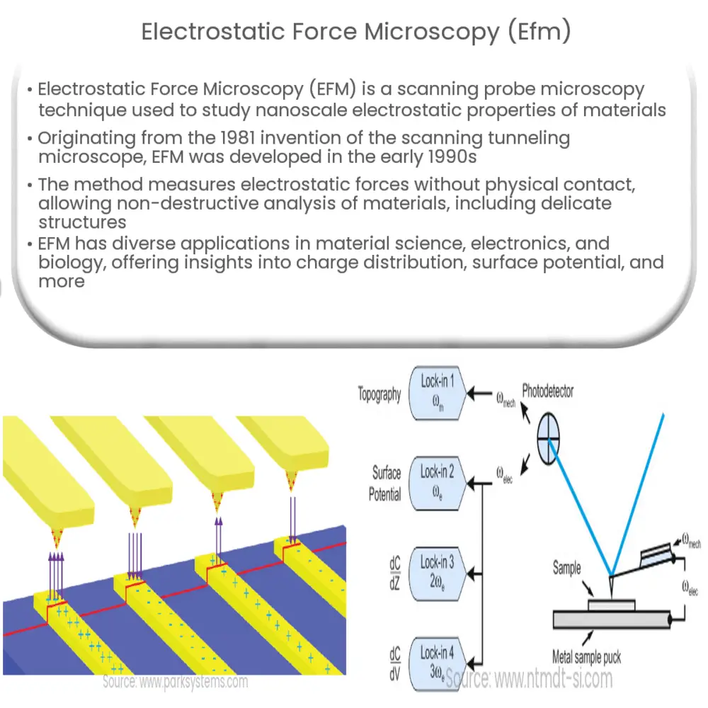
Electrostatic Force Microscopy (EFM) is an advanced scanning probe microscopy (SPM) technique used to investigate the electrostatic properties of materials at the nanoscale. By providing a direct, non-destructive method to visualize and quantify electrostatic interactions, EFM has become an indispensable tool for researchers working in various fields such as material science, electronics, and biology.
A Brief History of EFM
The development of EFM can be traced back to the invention of the scanning tunneling microscope (STM) in 1981 by Gerd Binnig and Heinrich Rohrer. They were awarded the Nobel Prize in Physics in 1986 for their pioneering work. The successful application of STM in imaging conductive materials at the atomic scale spurred the development of other SPM techniques, including atomic force microscopy (AFM) in 1986. EFM, a derivative of AFM, was first introduced in the early 1990s to explore the electrostatic properties of surfaces, with Martin, Williams, and Wickramasinghe being the first to demonstrate the technique.
Principles of EFM
At the core of EFM is the principle of measuring the electrostatic force between a sharp conductive probe and the sample surface. A bias voltage is applied between the probe and the sample, resulting in an electrostatic force between them. This force depends on the surface potential, the probe-sample capacitance, and the bias voltage. By scanning the probe over the sample and recording the force as a function of position, researchers can generate a spatially resolved map of the electrostatic properties of the sample.
EFM operates in a non-contact mode, which means that the probe does not make physical contact with the sample surface. This prevents damage to the sample and allows for the measurement of delicate structures such as thin films and biological samples. To maintain a constant distance between the probe and the sample, a feedback loop is used, which adjusts the probe’s position based on the changes in the electrostatic force detected by the probe.
EFM Applications
EFM has found a wide range of applications in various fields, including:
- Material Science: EFM can be used to study charge distribution, surface potential, and dielectric properties of materials such as polymers, semiconductors, and ferroelectric materials.
- Electronics: In the electronics industry, EFM is employed to investigate semiconductor devices, analyze failure mechanisms, and evaluate the performance of electronic components such as capacitors and transistors.
- Biology: EFM has been used to study the electrostatic properties of biological samples, including cell membranes, DNA, and proteins, providing insights into their structure, function, and interactions.
In conclusion, Electrostatic Force Microscopy is a powerful technique that offers unique insights into the electrostatic properties of materials at the nanoscale. Its non-destructive nature and high spatial resolution have made it an indispensable tool in a diverse range of research fields.
Advantages and Limitations of EFM
EFM has several advantages over other characterization techniques, including:
- Non-destructive: As a non-contact method, EFM can investigate delicate and soft materials without causing damage or altering their properties.
- High spatial resolution: EFM can achieve a resolution in the nanometer range, allowing researchers to study electrostatic properties at the nanoscale.
- Quantitative analysis: EFM can provide quantitative information about surface potential, charge distribution, and dielectric properties, helping researchers gain a deeper understanding of the materials being studied.
However, EFM also has some limitations:
- Tip-sample distance: Maintaining a constant distance between the probe and the sample is critical for accurate measurements, but this can be challenging due to variations in the sample’s topography.
- Signal interpretation: EFM signals are affected by various factors, including probe geometry, sample roughness, and environmental conditions, which can complicate data interpretation.
- Conductive samples: EFM is most effective when studying insulating or semi-insulating materials, but can be less accurate when characterizing highly conductive materials.
Recent Advances and Future Perspectives
Recent advances in EFM have focused on improving its performance, versatility, and ease of use. Some notable developments include:
- High-speed EFM: Novel scanning techniques and hardware have enabled faster imaging speeds, allowing researchers to study dynamic processes in real-time.
- Multi-frequency EFM: By using multiple frequencies simultaneously, researchers can obtain more information about the sample’s electrostatic properties, enabling a deeper understanding of the underlying physics.
- Integration with other techniques: EFM can be combined with other SPM techniques, such as Kelvin Probe Force Microscopy (KPFM) and Magnetic Force Microscopy (MFM), to provide a more comprehensive characterization of materials.
As EFM continues to evolve, it is expected that its applicability and capabilities will expand even further. Potential future developments may include:
- Improved resolution: Advances in probe technology and scanning techniques could enable EFM to achieve even higher spatial resolutions, possibly approaching the atomic scale.
- Expanded material compatibility: The development of specialized probes and methodologies could make EFM more effective for studying a wider range of materials, including highly conductive and magnetic samples.
- Environmental control: The integration of EFM with controlled environments, such as vacuum chambers or humidity-controlled cells, could help minimize the influence of external factors and improve measurement accuracy.
In conclusion, Electrostatic Force Microscopy is a versatile and powerful tool for investigating the electrostatic properties of materials at the nanoscale. As technology and methodologies continue to advance, EFM will undoubtedly play an increasingly important role in the research and development of materials and devices across a wide range of applications.